TomoWave Laboratories, Inc.
Proprietary medical imaging based on combined 3D opto...

Private Fundraise
This company may be interested in raising funds from accredited investors. You must Request Access to see more information about this company.
Request Access 9 TomoWave Laboratories:
Fast Facts
TomoWave Laboratories is commercializing proprietary biomedical imaging devices based on combined 3D optoacoustics and ultrasound invented by the founder.
We have developed imaging systems that show quantitative functional and molecular information on top of anatomical tissue structures. This provides comprehensive medical information for accurate detection, diagnostics, image-guided surgery and thermal therapy of cancer and vascular diseases.
Groundbreaking
Shift in Diagnostics
 Diagnostics has come a long way, but it hasn’t come far enough.
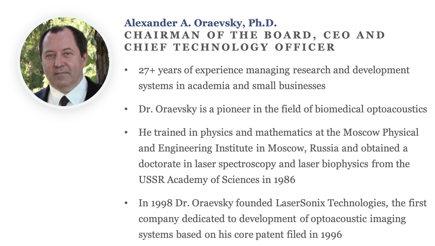
Wanting to see the inside of a human body with his own eyes, Dr. Alexander Oraevsky recognized this problem firsthand. He knew that other doctors would also like to take a deeper look and hopefully detect and diagnose diseases more quickly and efficiently.
For many, this might seem like fantasy. It is presently impossible to see under the skin because light scatters in human tissues, making even highly contrasted objects, such as blood vessels, blurry. But Dr. Oraevsky's work has led to a solution!
Seeing by Listening to
the Sound of Light
 In the early 1990s Alexander worked on a project of precise laser microsurgery. He used short laser pulses for ablation (removal) of biological tissue, and he did so without thermal damage to adjacent tissue. He heard sound when laser pulses interacted with tissue, so he decided to detect this sound with pressure detectors and measure the spectrum of this sound in order to get feedback information from the process of pulsed laser ablation.
He quickly realized that the acoustic signal amplitude was much greater outside of the audible range of sound, called ultrasound. Ultrasound signals from blood vessels and other biological tissues with strong absorption of light were very intense even with dramatically reduced energy of laser pulses. The “aha!" moment came when, during an experiment made together with his research associate, Rinat Esenaliev, Dr. Oraevsky could detect an acoustic signal from a small piece of liver deeply embedded within a stack of chicken breast tissues.
This led to the invention of the depth-resolved optoacoustic imaging method, which allows one to see in the depth of tissues with high spatial resolution by listening to the sound of light absorbed in the body.
Now TomoWave Laboratories is developing and commercializing a platform technology of three-dimensional optoacoustic tomography for detection and diagnostics of cancer and vascular diseases.
Now TomoWave Laboratories is developing and commercializing a platform technology of three-dimensional optoacoustic tomography for detection and diagnostics of cancer and vascular diseases.
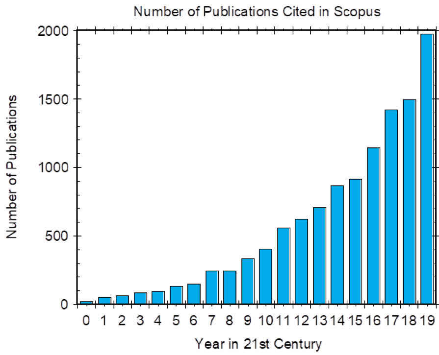 In 2000 Dr. Oraevsky founded an annual conference under auspices of International Society for Optical Engineering (SPIE) to discuss the latest advances in his technology. It has quickly become the largest conference within 20,000 attendees Photonics West Symposium. This conference stimulated exponential growth of the research community and optoacoustic imaging has become the fastest growing biomedical imaging technology in the 21st century (see plot of the number of publications in 21st Century in the area of optoacoustic/photoacoustic imaging).
First Commercial Product
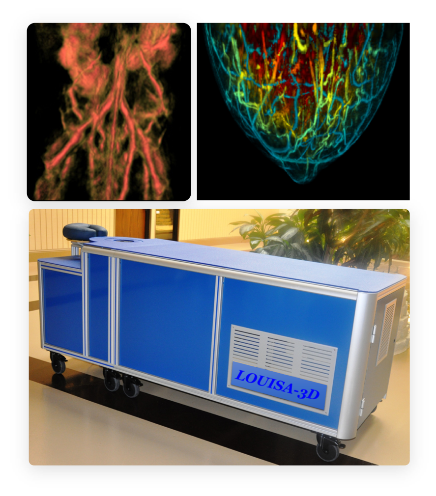
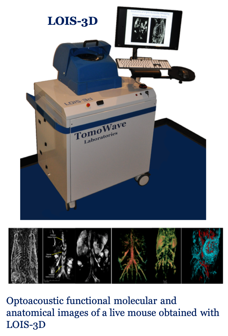 In response to a large demand from biomedical researchers to have possibility of high resolution visualization of diseases through entire body and brain of mouse models, TomoWave developed and in the end of 2019 launched sales of Laser Optoacoustic Imaging System (LOIS-3D).
While standard systems (Xray, MRI, Ultrasound) provide only anatomical images, but neither functional info on blood content and oxygen saturation nor highly resolved molecular composition. Fluorescence and bioluminescence systems provide images with poor resolution.
LOIS3D is a full view tomographic system provides info to researcher about anatomy, tissue function and molecular composition with unmatched tissue contrast and 0.25 mm resolution equal in all 3 dimensions.
A Major Problem
that needs to be solved
Breast cancer is a horrific, devastating disease that is so evasive and so difficult to detect that some women actually surgically remove their breasts as a precautionary solution.
Genetics play a major role in that decision, no doubt. But the weak diagnostics solutions aren't helping the situation.
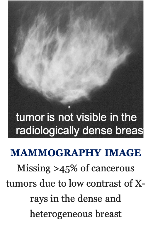
The primary breast cancer screening consists of a mammography based on the ionizing radiation of X-rays.
Breast density plays a factor in the success of these tests, so younger women are not great candidates. And on average, mammography misses 25% of all cancerous lesions. This is worse in the dense heterogeneous breast -- i.e., greater than 50,000 of breast cancer cases in United States only, and more than 100,000 breast cancers per year worldwide.
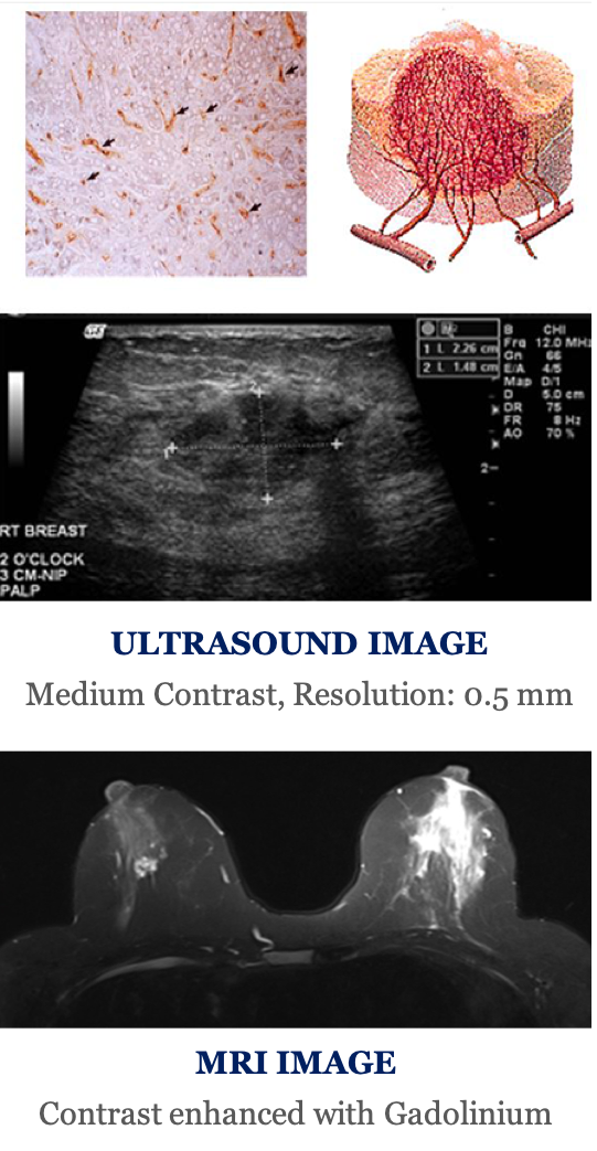
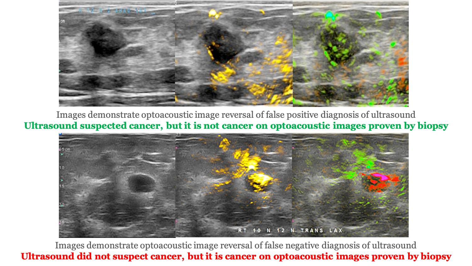
Clinicians use ultrasound imaging as an additional tool to improve cancer detectability, and patients undergo breast biopsy or surgery for definitive diagnosis. Ultrasound significantly improves detection sensitivity to 95-97%, thus reducing the number of false negative diagnoses. However, ultrasound cannot visualize tumor micro vasculature – hallmark of aggressive cancer. Low specificity of breast tumor differentiation by ultrasound results in 7-8 negative biopsies out of every 10 performed.
This is what the medical field refers to as being "over diagnosed," to the tune of 1 million unnecessary biopsy procedures every year in the US. These unnecessary procedures total approximately $3 billion. In the European Union there are more than 500,000 negative biopsy procedures, with costs of about $700 per procedure. These unnecessary procedures may cause pain, severe anxiety and sometimes infection.
MRI and PET/CT modalities use toxic contrast agents for breast cancer detection, take about 45 min, and are often unaffordable to smaller hospitals and patients.
One Groundbreaking
Solution
TomoWave Laboratories is solving these problems by commercializing proprietary biomedical imaging devices based on combined 3D optoacoustics and ultrasound invented by the founder.
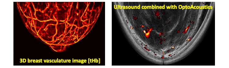
Optoacoustic tomography provides for noninvasive means to determine physiological conditions of the tumor angiogenesis (density, leakiness of microvessels and hypoxic state) independently on breast density.
Optoacoustic imaging and monitoring technologies (that combine light and sound to achieve high-contrast and high-resolution visualization and quantitative characterization of biological tissues) represent a very lucrative commercial opportunity.
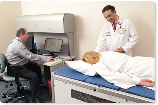
 Dr. Oraevsky, founder of TomoWave, is the pioneer and inventor of the optoacoustic imaging method and system. The commercialization of the laser optoacoustic ultrasonic imaging system (LOUISA-3D) can substantially improve existing screening (detection) and diagnosis, reducing the adverse psychological and financial impact of performing unnecessary breast biopsies and ineffective chemotherapy treatments.
Moreover, LOUISA-3D has the potential to improve early detection of breast cancer, especially in younger women with dense breasts, by providing coregistered anatomical and functional information of the lesion.
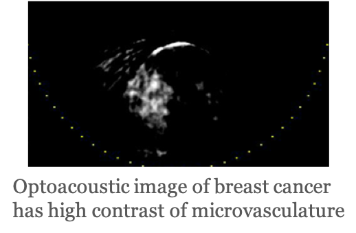
The optical contrast of LOUISA is based on oxygenated and deoxygenated hemoglobin of blood in tumor angiogenesis. The ultrasonic contrast images are obtained by LOUISA using B-mode ultrasound, which provides high sensitivity and resolution for visualization of the dense core of malignant breast tumors.

Malignant tumors have high concentration of microvasculature, hypoxic blood, uneven shapes and greater speed of sound, allowing for noninvasive clinical distinction between benign masses and malignant tumors using optoacoustic images coregistered with B-mode ultrasound.
2D optoacoustic functional images were proven to improve diagnostic specificity of ultrasound two-fold (Oraevsky et al. Photoacoustics 2018, DOI: 10.1016/j.pacs.2018.08.003). Automatic 3D system is designed to perform even better providing quantitative functional maps within anatomical tissue structures.
Vasculature and microvasculature of tumors are important medical factors
Optoacoustic imaging visualizes vascular and microvascular supply of the tumors and measures functional changes at the molecular level enabling more accurate diagnostics. It is expected that optoacoustic images may be able to detect early physiological changes in the tumor indicating whether therapy is effective or not.
LOUISA can fulfill the market need for an affordable imaging modality with high sensitivity and high specificity for breast cancer (other types of cancer can be detected with the next generation modalities). This is one of many disease applications within LOUISA platform technology.
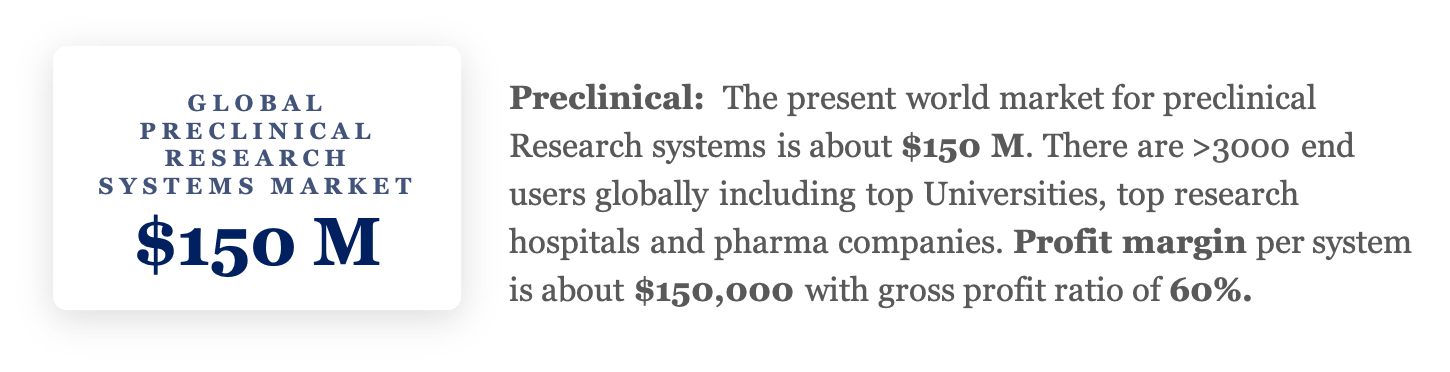
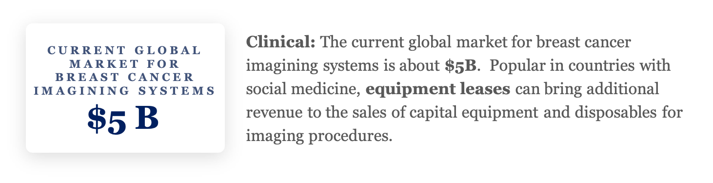
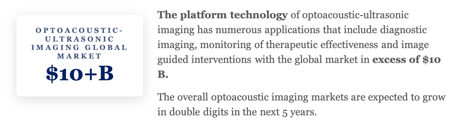
Markets for the
Platform Technology
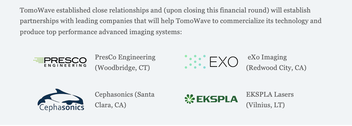
Traction & Accomplishments
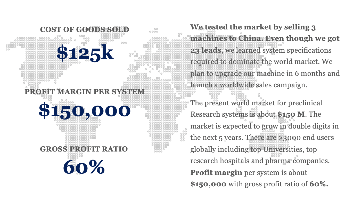
Three LOIS-3D systems have been purchased by University Laboratories. Bids for these systems have been won through comparative performance analysis by an independent purchasing committee.
Five premiere hospitals in the US, Europe, Asia, and Israel have expressed interest in purchasing our systems for breast cancer protection. These hospitals include:
- The University of Texas MD Anderson Cancer Center, Houston, Texas
- Moores Cancer Center, University of California San Diego, California
- Karmanos Cancer Institute, Wayne State University, Detroit, Michigan
- Breast Cancer Care Unit, Assaf Harofeh Medical Center, Tel Aviv, Israel
- The Third Affiliated Hospital, Sun Yat-Sen University, Guangzhou, China
Twenty-three leads for future system sales have been acquired through demonstration of better specifications and performance compared with the competition.
Dr. Oraevsky also holds seven core patents behind the technology.
Meet The Team